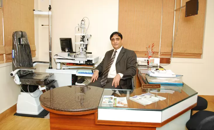
Doctor Eye Institute, established in 1964, is a state of art eye care center with all the latest and most modern ophthalmic equipment and facilities. For the last 60+ years in service, Doctor Eye Institute Pvt. Ltd. has been the pioneer in providing many world-class treatments for Total Eye Care in Mumbai, India. For decades we have provided the best equipment and facilities keeping updated with changing times and technology – a pursuit for excellence has been our tradition.
An ISO 9001 – 2008 Certified and NABH Certified hospital with the following facilities.
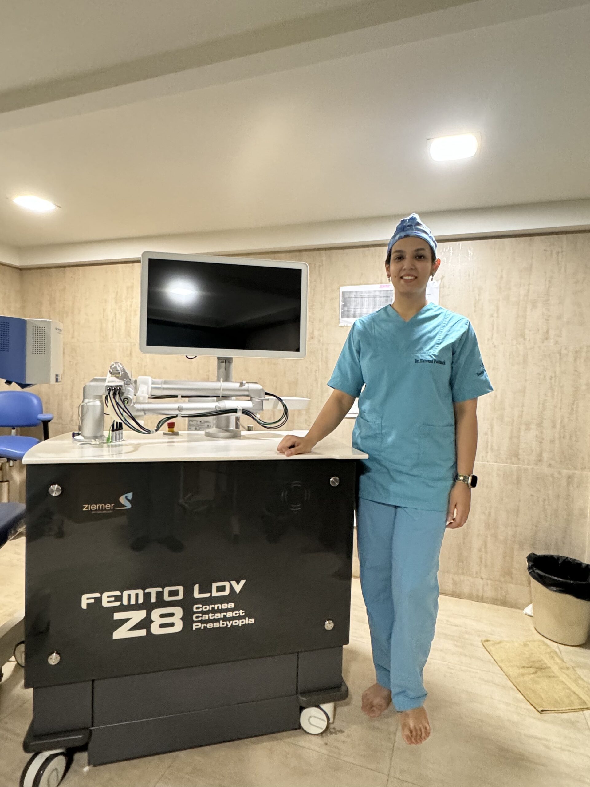
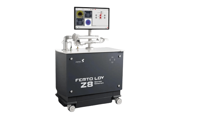
Robotic Laser Bladeless Cataract surgery by Phaco emulsification using Femto Laser Z8 and White star Signature machine. (This machine is first in Maharashtra and third in whole of India.)
LASIK surgery for correction of refractive error or removal of glasses using ESIRIS SHWIND EXCIMER LASER WITH ONLINE PACHY.
Glaucoma diagnostic and surgery equipment’s.
Equipment’s for evaluation and treatment of Retinal Disorders like FFA, B scan, Green laser, Vitrectomy etc.
Facility for performing specialized surgeries like Squint, Keratoplasty or Corneal Grafting, Oculoplastic Surgeries etc.
FacilitiesavailableatDoctorEyeInstitute
Complete Eye Checkup and Screening.
Advanced Femto Laser Robotic Bladeless Cataract Surgery and Phaco emulsification setup with IOL implantation (foldable Monofocal/Multifocal and TORIC lenses)
Specular Microscopy
Glaucoma Screening and Surgeries . (Perimetry, Gonioscopy, Non Contact Tonometer, Trebeculoctomy and Express Shunts implants)
CLE/ PHAKIC Refractive Lenses (PRL) implantation where Lasik is not possible
Retinal Surgery Setup. (F.F.A. with Digital Imaging , B Scan, Colour Photographs, OCT)
Yag Laser for after Cataract and Glaucoma Surgeries
Cornea Grafting, C3R (keratoconus Treatment)
Oculoplasty. (Plastic Surgery around the eye)
Squint Operations
Spectacles & Contact Lens Clinic
Non Contact Tonometer
Purpose: Measures intraocular pressure (IOP) without touching the eye.
How It Works: Uses a puff of air to flatten the cornea and measure the resistance, correlating with eye pressure.
Benefits: Quick, painless, and essential for glaucoma screening. It avoids discomfort and potential risk of infection associated with contact tonometry.
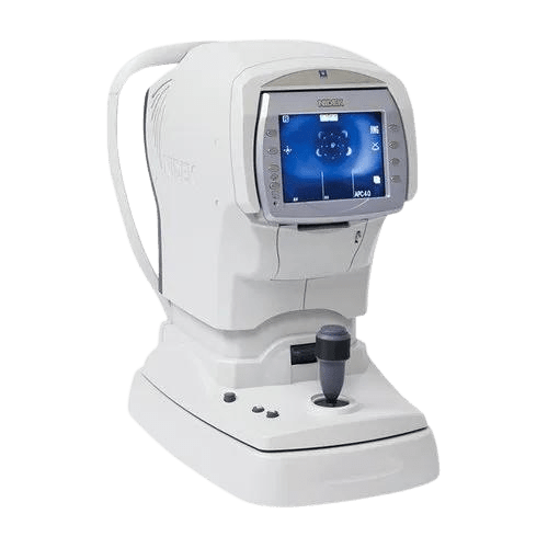
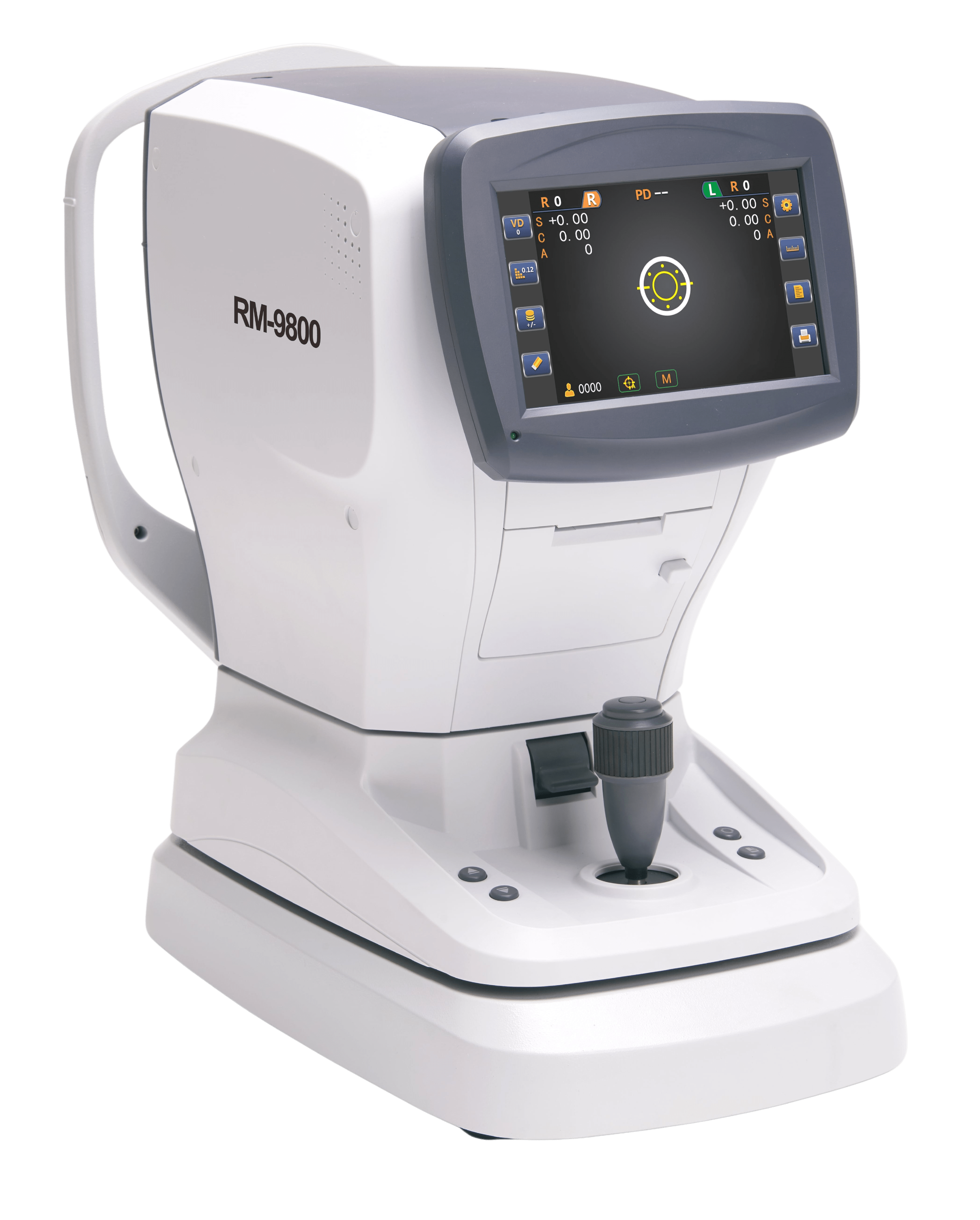
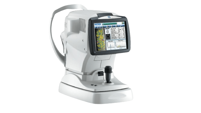
Auto Refractometer
Purpose: Determines the refractive error of the eyes.
How It Works: Automatically measures how light is refracted through the eye, providing precise measurements of nearsightedness, farsightedness, and astigmatism.
Benefits: Provides accurate and quick measurements for prescribing glasses or contact lenses, enhancing the efficiency of eye exams.
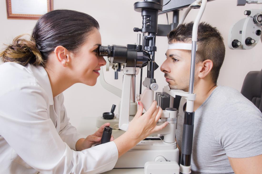
Slit Lamp
Purpose: Examines the anterior segment of the eye, including the cornea, iris, and lens.
How It Works: Uses a high-intensity light source and a microscope to illuminate and magnify the eye's structures.
Benefits: Allows for detailed examination of the eye’s anatomy, aiding in diagnosing various conditions such as cataracts, macular degeneration, and corneal ulcers.
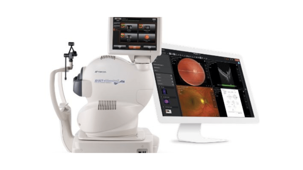
Optical Coherence Tomography (OCT)
Purpose: Provides detailed cross-sectional images of the retina and optic nerve.
How It Works: Uses light waves to capture detailed images of the retina’s layers, helping detect and monitor diseases like glaucoma, diabetic retinopathy, and macular degeneration.
Benefits: Non-invasive and painless, offering high-resolution images essential for early diagnosis and effective treatment planning.
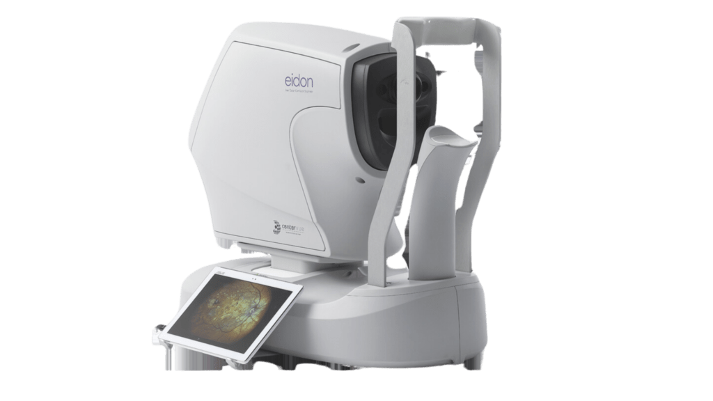
Fundus Camera (EDION Wide-Field Retinal Imaging)
Purpose: Captures detailed images of the retina.
How It Works: The EDION Fundus Camera can capture retinal images up to 220 degrees without the need for pupil dilation.
Benefits: Provides comprehensive retinal assessments, covering a vast area of the retina in a single image, crucial for detecting peripheral retinal diseases.
ANTERION
Purpose: Multifunctional imaging for anterior segment analysis.
How It Works: Uses high-resolution swept-source OCT technology to provide detailed imaging and biometry of the anterior segment, including the cornea, anterior chamber, and lens.
Benefits: Offers a comprehensive analysis of the anterior segment for diagnosing and managing conditions such as keratoconus, cataracts, and glaucoma. Enhances surgical planning and postoperative assessment with detailed and accurate measurements.
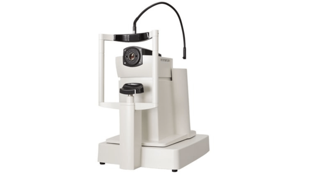
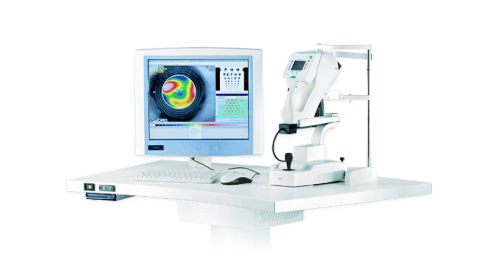
Pentacam Topographer
Purpose: Provides comprehensive corneal topography and tomography.
How It Works: Uses a rotating camera to capture multiple images of the cornea, creating a detailed 3D map of its shape and thickness
Benefits: Essential for diagnosing keratoconus, planning refractive surgery, and assessing corneal health.
Corneal and Ocular Wavefront Machines
Purpose: Measures aberrations in the eye to improve visual outcomes.
How It Works: Analyzes how light waves travel through the eye, identifying irregularities that affect vision.
Benefits: Helps in customizing treatments for refractive surgery, leading to better visual outcomes and reduced higher-order aberrations.
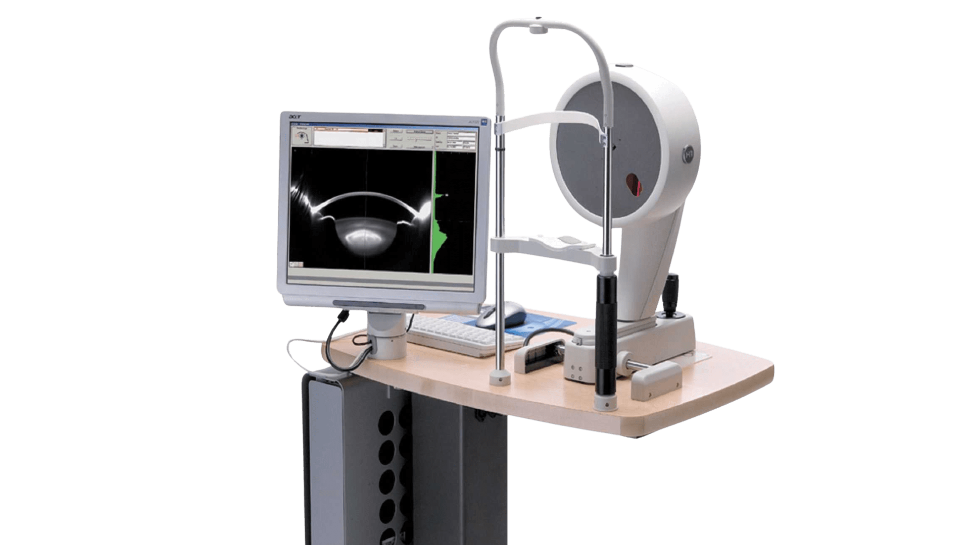
Specular Microscope
Purpose: Examines the endothelium, the innermost layer of the cornea.
How It Works: Captures detailed images of the corneal endothelial cells.
Benefits: Assesses endothelial cell health, crucial for diagnosing corneal diseases and planning surgeries like corneal transplants.
TreatmentEquipments
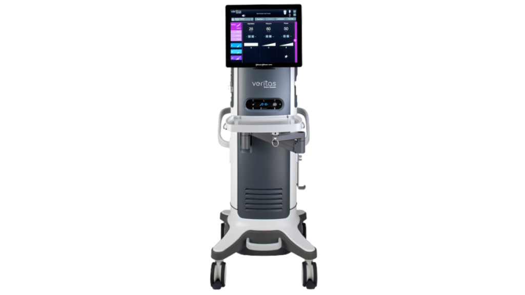
Veritas Phaco Machine
Purpose: Used for cataract surgery.
How It Works: Utilizes ultrasound technology to emulsify the cloudy lens, which is then removed and replaced with an artificial intraocular lens.
Benefits: Advanced fluidics and real-time occlusion sensing enhance safety, efficacy, and accuracy. Reduced heat generation minimizes thermal injury, leading to quicker recovery and better visual outcomes.
Laser Systems for Refractive Surgery (LASIK and CLEAR)
Purpose: Corrects refractive errors like myopia, hyperopia, and astigmatism.
How It Works: LASIK uses a laser to create a corneal flap and reshape the underlying corneal tissue. CLEAR involves creating a small lenticule within the cornea, which is then removed through a tiny incision.
Benefits: Both methods are highly precise, minimally invasive, and offer quick recovery times with excellent visual outcomes.
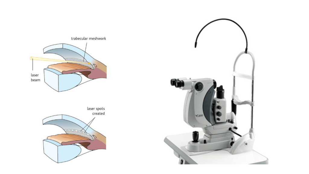
Selective Laser Trabeculoplasty (SLT)
Purpose: Treats glaucoma by reducing intraocular pressure.
How It Works: A laser selectively targets the trabecular meshwork, enhancing fluid outflow from the eye.
Benefits: Non-invasive, repeatable, and effective in lowering eye pressure with minimal discomfort.
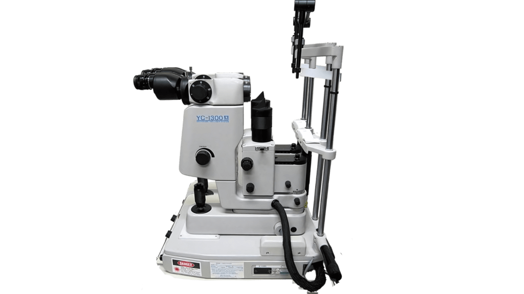
YAG Laser Capsulotomy
Purpose: Treats posterior capsular opacification (a common post-cataract surgery complication).
How It Works: Uses a YAG laser to create an opening in the cloudy capsule that surrounds the intraocular lens.
Benefits: Quick, painless, and restores clear vision effectively.
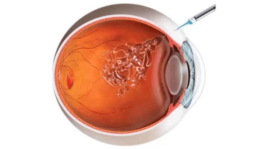
Intravitreal Injection Systems
Purpose: Delivers medication directly into the vitreous cavity of the eye.
How It Works: Injections are used to administer anti-VEGF drugs or steroids for conditions like diabetic retinopathy and macular degeneration.
Benefits: Provides targeted treatment with high efficacy in reducing retinal swelling and inhibiting abnormal blood vessel growth.
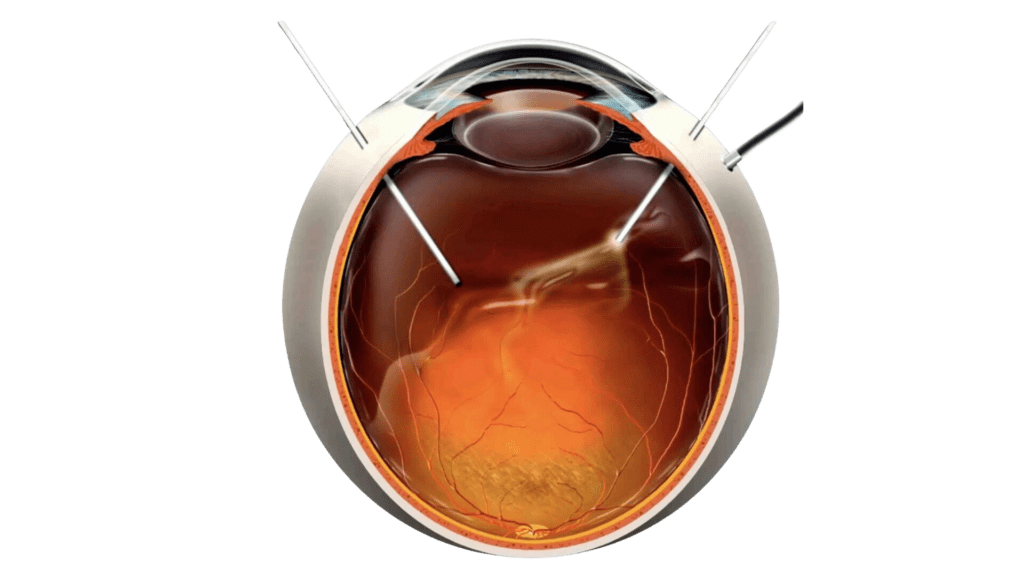
Vitrectomy Machine
Purpose: Used for vitreoretinal surgery to treat conditions such as retinal detachment, macular holes, and diabetic retinopathy.
How It Works: Removes the vitreous gel and replaces it with a saline solution or gas bubble to allow the retina to heal.
Benefits: Restores lost vision, prevents further retinal damage, and treats various vitreoretinal disorders effectively.
Best Eye Hospital in Mumbai for Lasik, Cataract, Retina, Glaucoma and Laser Eye Surgery.